Seminar: Van Thompson, November 10, 2014
CISMMS Special Seminar
Dental Enamel: A Multi-Scale Modelling Challenge
Van Thompson PhD, DDS
Professor of Biomaterials, Biomimetics, & Biophotonics
King’s College London Dental Institute
Floor 17, Tower Wing, Guy’s Hospital
London, SE1 9RT, United Kingdom
The tooth is a unique functionally graded composite structure at several levels providing a hard and apparently self-healing external enamel structure bonded to a dynamic and resilient dentin core both supported by a vascular and neural network in the tooth pulp. Tooth enamel is nature’s cell-derived method for production of a high elastic modulus (~ 90 GPa), hard, wear, and fatigue resistant structure. Enamel is built from prisms/rods each laid down by a single ameloblast cell. The enamel prism nanostructure is a generally well characterized. Each interlocking prism (~ 5 µm dia) is primarily composed of nanocrystalline carbonated hydroxyapatite (CHA) with orientations that provide graded properties. The arrangement of the prisms as the ameloblast grow outward from the dentoenamel junction (DEJ) is termed decussation. Although not well characterized it is known that bundles of 50-100 prisms course outward at + and – 40° from the radial direction in alternating layers (light and dark bands in Figure) that are roughly parallel to the occlusal plane of the tooth. Locally these layers are thought to undulate in an occlusao-gingival direction. In addition the overall layer direction moves occlusally as it develops outward to what becomes the surface of the enamel. This is known as multi-serial decussation as bundles of prisms change course while in the thin enamel of mammals such as rats serial decussation predominates where single cells form complex intertwining patterns. These structures have graded properties. In humans, prism deccusation provides enamel with an increasing fracture toughness (R curve behavior with KIC increasing from 0.5 to 2.4 MPa·m0.5) whether cracks are initiated at the enamel surface or from the DEJ. The generally planar nature of the decussation allows enamel to crack from the incisal or occlusal to gingival but this vertical cracking of teeth does not lead to loss of enamel, failure of the structure, or caries (tooth decay). Enamel decussation is extensive at tooth cusps where space filling is more difficult (histologic investigation refer to this as gnarled enamel). Unique to all enamel outer surfaces is the parallel alignment of prisms with little evidence of decussation. This goes counter to the belief that one ameloblast creates one prism and that prisms course from the DEJ to the surface and do not change diameter. The surface area of the enamel is roughly 50% higher than the surface area of at the DEJ where ameloblasts align to begin enamel development. To date there have been no investigations of enamel structure to determine the nature and extent of decussation in 3 dimensions.
Critical to the support and maintenance of the enamel is dentin. Dentin is approximately 60% by weight CHA making it similar to bone and has an elastic modulus that ranges from 12-18 GPa. The dento-enamel junction (DEJ) is the transition zone between enamel and dentin with a width of 50-100. This graded interfaces surface plays a role in limiting fracture initiation at the DEJ. This presentation with review studies on the Hertzian contact elastic response and Vickers indentation at high strain rates of enamel and dentin. These finding will be related to fracture toughness measurements of enamel and dentin by several groups and the need to further explore the graded structure mechanical response with enamel location and orientation in the tooth. Emphasis well be on the role of decussation on enamel properties and likely mechanisms for the repair of enamel microcracks that originate at the enamel surface or near the DEJ when teeth are fatigued.
About the Speaker
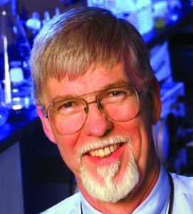
Van P Thompson, DDS, PhD, is currently, Professor of Biomaterials, Biomimetics and Biophotonics at King’s College London Dental Institute and was previously Chair, Biomaterials and Biomimetics, NYU College of Dentistry. Known for his work on adhesion and bonded bridges at the University of Maryland he has published many articles and made numerous presentations on dental biomaterials in the U.S. and internationally. His current research areas include dentin caries activity, all-ceramic crown fatigue and fracture, modifications of dentin for bonding, engineering tissue response via scaffold architecture and practice based research (PEARL Network).


