
Quantitative voxel-to-voxel comparison of TriBeam and DCT strontium titanate three-dimensional data sets
In this article, time-resolved (four-dimensional) and high-resolution data have been collected using X-ray diffraction contrast tomography (DCT) and the TriBeam from the same volume in a single strontium titanate sample. Methods presented for the merger of the reconstructions enable direct quantitative comparisons on a grain and voxel scale and can be extended to other multi-modal data sets.
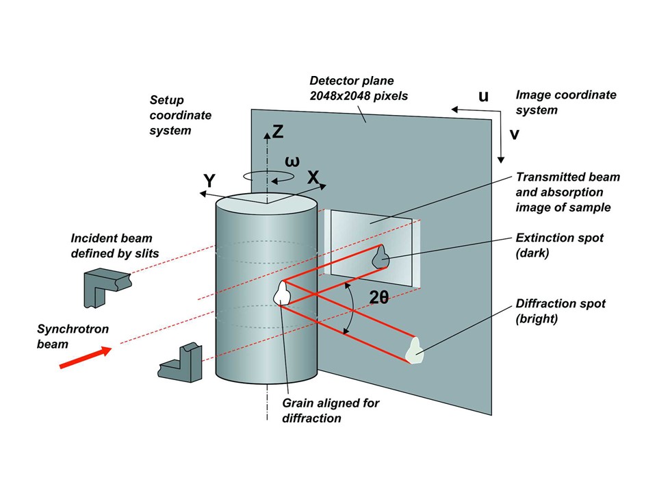
X-ray DCT experiments are performed by irradiating a sample with a beam defined by apertures or slits in the beamline. The sample is rotated in increments of ω = 0.05°, around the z axis. Extinction and diffraction spots simultaneously appear and disappear on the transmitted-beam region of the detector (center of detector) and in the surrounding area (diffraction spots). Figure reproduced after Ludwig, Reischig et al. JOURNAL OF APPLIED CRYSTALLOGRAPHY, Volume 48| Part 4| June 2015| Pages 1034-1046; doi:10.1107/S1600576715009231
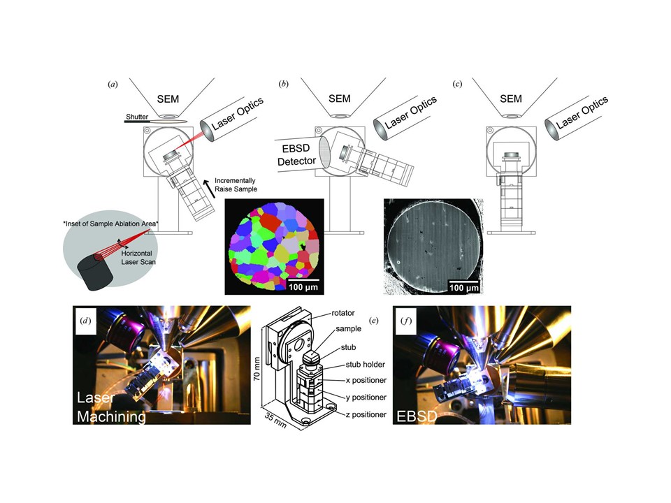
TriBeam experiments use a high-precision piezoelectric stage to position the sample within the microscope chamber. (a) The sample is tilted to 30° for surface parallel femtosecond laser ablation. The laser is scanned horizontally across the sample surface to remove the material which has been incrementally raised into the focused beam path. (b) The piezoelectric stage is tilted for EBSD data collection (70°) with the detector inserted. (c) The sample is tilted to 0° for SEM imaging. Photographs of configurations (a) and (b) are shown in (d) and (f), respectively. (e) A schematic of the piezoelectric stack with components labeled is shown. Courtesy of JOURNAL OF APPLIED CRYSTALLOGRAPHY Volume 48| Part 4| June 2015| Pages 1034-1046; doi:10.1107/S1600576715009231